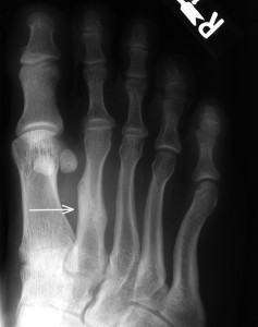
Stress fractures of the foot and lower extremity are not uncommon. Mostly they occur in sports people, athletes and people with high demand occupations which require long periods of weight bearing.
A stress fracture is a ‘crack’ in the bone – usually the outside layer (the cortex). In the foot, they can occur in metatarsals, and less commonly the navicular, cuboid and calcaneus bones – however any bone has the potential to develop a stress fracture given the right circumstances.
The most common location for stress fractures in the foot is within the 2nd and 3rd metatarsal bones – as they can tend to take a lot of weight bearing force in certain foot types. Most patients with complain of aching and swelling on top of the forefoot, which is exacerbated by extended periods of weight bearing or sports.
Usually a stress fracture is easy to diagnose with a plain x-ray. However, in the early stages (the first week or two), x-rays may not clearly demonstrate the problem, and other types of imaging might be recommended (eg MRI or bone scan).
It is important to remember that stress fractures will heal if rest, activity modification, and offloading the area are done. However, sometimes we also need to immobile the foot and ankle in a removable cast walker if it is difficult to avoid using the area due to work or training commitments. If there is a suspicion there is an underlying problem with your bone density (strength), we will often recommend a bone mineral density test to check for osteopaenia or osteoporosis via your GP, and a check on your vitamin D and calcium levels.


